
The results verify that deep learning-based classifiers trained on MIDOG and ATYPIA have difficulties to recognize mitosis on our dataset which shows that the created mitosis dataset has unique features and characteristics. Various deep learning classifiers and the proposed classifier are trained with a publicly available mitosis datasets (MIDOG and ATYPIA) and then, validated over our created dataset. In the classification part, the detected isolated nucleus images are passed through proposed MITNET-rec deep learning architecture, to identify the mitosis in the WSIs. In MITNET-det architecture, to extract features from nucleus images and fuse them, CSPDarknet and Path Aggregation Network (PANet) are used, respectively, and then, a detection strategy using You Look Only Once (scaled-YOLOv4) is employed to detect nucleus at three different scales. The created datasets are used to train the MITNET network, which consists of two deep learning architectures, called MITNET-det and MITNET-rec, respectively, to isolate nuclei cells and identify the mitoses in WSIs. The first dataset is used to detect the nucleus in the WSIs, which contains 139,124 annotated nuclei in 1749 patches extracted from 115 WSIs of breast cancer tissue, and the second dataset consists of 4908 mitotic cells and 4908 non-mitotic cells image samples extracted from 214 WSIs which is used for mitosis classification. Moreover, this paper introduces two new datasets. In this paper, a two-stage deep learning approach, named MITNET, has been applied to automatically detect nucleus and classify mitoses in whole slide images (WSI) of breast cancer. The inter, and intra-observer variability of this assessment is high.

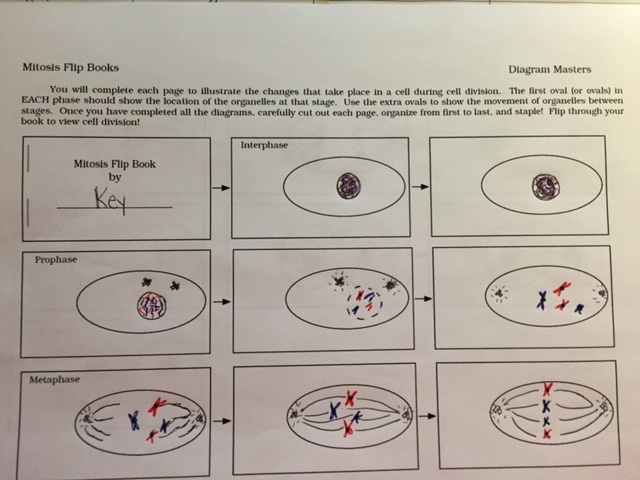
Well-rendered visuals that enhances product & aids understanding.Mitosis assessment of breast cancer has a strong prognostic importance and is visually evaluated by pathologists. _ visible and correct phase abbreviation on each page.25 pages = 5 per phase (PMAT + cytokinesis) _ spindle fibers appear / location / disappearĮxemplary, neat, well planned format that enhances product._ centrioles appear / location / disappear._ (2) chromosomes appear / location / disappear.AttributeĪccurate, complete, extensive information. Your own computer-generated images or templates are acceptable, but check with the instructor. Each stage will have at least five pages.

Illustrate the four phases of mitosis AND include cytokinesis.

A flip book is a way to do a cartoon animation showing gradual change on each page. You will create a flip book to illustrate the stages of cell mitosis (not the cell cycle nor meiosis).


 0 kommentar(er)
